The Stone Service
Facts and figures
Nephrolithiasis (stone formation in the kidneys) is a recurrent condition with significant morbidity. While symptomatic episodes require appropriate treatment, prophylactic work up to prevent recurrence is of great importance.
- Kidney stones are found in as many as 1 in 10 people
- Kidney stones most often affect people aged 30-60
- Stones are more common in men than women
What causes stones?
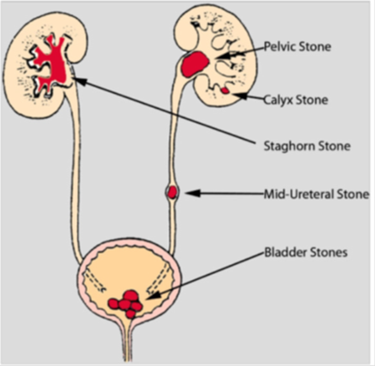
The function of the kidneys is to remove waste products from the blood. If the concentration of waste products is significantly increased, the waste products can crystallise in the kidneys and eventually form hard stones. Stones can form anywhere along the urinary tract.
Kidney stones can be caused by a variety of things, including dehydration, diet, medications or infections. Rare genetic conditions that predispose to stone formation include primary hyperoxaluria and cystinuria amongst others.
Recurrence of kidney stones is very common but can be easily prevented with appropriate investigation and management. Lifestyle and dietary advice can help to reduce the risk of recurrence.
Treatment
Treatment may consist of surveillance, breaking up the stones using shock waves (lithotripsy) or surgery with ureteroscopes where the stones are broken up with a laser. These do not involve any surgical cuts to the skin. For larger stones, a telescope can be placed into the kidney remove the stone. This is known as percutaneous nephro-lithotomy or PCNL. Open surgery is very rarely needed. Further information about the treatment of stones, can be found on Guy’s and St Thomas’ NHS Foundation Trust website:
http://www.guysandstthomas.nhs.uk/our-services/urology/specialties/stone-unit/patients.aspx
Why analyse stones?
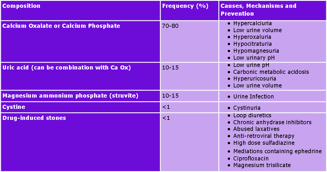
Determination of the chemical composition of stones aids investigation of the pathophysiology of stone formation, and will help to decide treatment modality and prevent recurrence.
How do we analyse stones?
At Viapath, stones are analysed using fourier-transformed infrared (FT-IR) spectroscopy, which is a specific, rapid and versatile method. Infra-red radiation causes atomic vibrations and consequently energy absorption, which results in different absorption bands for different chemical compositions. Scans are then interpreted using in house calibration models and spectral libraries.
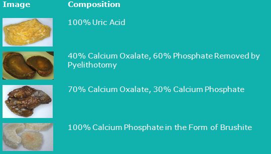
Images of kidney stones passed or removed from patient
How can we help?
The Stone Unit based at Guy’s Hospital is one of the largest in the country. It has state of the art treatment options including a dedicated operating theatre for patients with stones. Viapath offers a stone analysis service in Reference Chemistry which is complemented by metabolic investigations in the Inherited Metabolic Disease Laboratory (urine amino acids for cystine, ornithine, arginine and lysine) and genetic/molecular investigations in the Purine Research Laboratory (APRT/HPRT mutations) within Biochemical Sciences. The Genetics Laboratory based at Guy's Hospital offers cystinuria genetics.
Stone samples in plain universal containers can be sent to Viapath’s Reference Chemistry Laboratory at St. Thomas’ Hospital. A detailed analysis report is provided and turnaround times are 7 working days. The laboratory can also be contacted for further clinical advice and guidance on the emails below.
Contact Details
Reference Chemistry Laboratory at St Thomas'
4th floor, North Wing, St Thomas' Hospital, Westminster Bridge Road, London SE1 7EH
Email: Zehra [dot] Arkir [at] viapath [dot] co [dot] uk
References
Brener, Z. Z., et al., 2011. Nephrolithiasis: Evaluation and Management. Southern Medical Journal, 104 (2), 133-139.
C.Turk, T Knoll, A.Petrik, K Sarica, C Seitz and M Straub, 2014. Guidelines on Urolithiasis. Click here [Accessed 29/10/2015]
NHS Choices, 2016. Kidney Stones. Click here [Accessed 18/01/17]
Evan, P, 2010. Physiopathology and etiology of stone formation in the kidney and the urinary tract. Paediatric Nephrology, 2010. 25 (5) 831-841.