The History Of Histological Dissection: Landmarks And Pointers Of The Past, Present And Future
A glance at the history books tells us a great deal. Let’s begin with the word “dissection” derived from the Latin “dissecare” meaning “to cut to pieces”. This is quite a blunt description of what we now understand by modern dissection however it reflects the key premise of the act to dissect.
Dissection was used primarily to explore and evaluate anatomy and later improve understanding of physiology. Early reports suggest that human dissection was carried out by Greek physicians in the early part of the 3rd century BC. The Romans also had a significant role to play in the development of medicine. The most widely recognised practitioner of the time being Galan (129 CE) who lectured, wrote and exhibited extensively on anatomy and the value of dissection as a learning tool.
Despite strong resistance to the practice of human dissection, the Greeks were keen to establish a hub of medical knowledge and the government of the time supported its practice. However histological dissection became a taboo subject around the globe. This was as a result of religious beliefs merged with fear and trepidation about what might happen to the preservation of the human soul if dissection was performed on corpses. This resistance continued in the UK until 1832 when the first Human Anatomy Act was passed. Up to then it was an outlawed practice and medical professionals of the time would go to great lengths to acquire the cadavers required to study human anatomy. This spawned the era of the “body snatchers”. The passing of the Act paved the way for the modern-day study of anatomy and physiology and gave a license to doctors, teachers of anatomy and bona fide medical students to dissect donated bodies.
What then have been the significant advances in terms of the practice and equipment used for medical practice and more specifically histological dissection? Again the Romans made significant strides forward in this area, with the development of a wide range of surgical instruments. The most widely known example is the scalpel. Made mainly from bronze or steel, arguably this is the most valuable dissection instrument for incisional and deep or long cutting of tissue and the almost identical form is retained in scalpels used today. Other examples include bone drills, bone forceps, male catheters, vaginal specula, spatulas and the surgical saw.
Advances in cellular pathology
Modern day dissection surprisingly has not progressed significantly over time. Our understanding of anatomy and physiology has progressed but our methods of assessing or evaluating macroscopic dissection procedures has relied mainly on surgical instruments used in the operating theatre, rather than improving on dissection instrumentation at the cut up bench. The discipline of cellular pathology has advanced tremendously and we have seen some very impressive improvements in the developing technologies. Some examples include the introduction of enclosed tissue processing equipment, sophisticated paraffin embedding machines, improved microtomes for precision in section cutting. Most significantly we have seen the rise of automation with the introduction of a plethora of staining machines for routine haematoxylin and eosin (H&E) staining, special staining and immunocytochemical (ICC) staining procedures and also have automated coverslipping machines. The growing developments in molecular technology and equipment also looks set to be an area of great expansion in the future.
We have, perhaps, spent a great deal of time perfecting and improving on new technologies without necessarily working out the parameters that ensure that the tissue we assess is optimal for the procedures we need to investigate. A classic case in point is the massive explosion of publications on the use of ICC in cellular pathology during the 1980s and 90s. There was great interest in developing automated platforms and also improving sensitivity for the detection of ever-smaller antigenic epitopes. However as we attempted to identify an ever-increasing panel of antibodies to work on paraffin sections recognition dawned that optimising tissue fixation and processing was more important to the final results than was originally believed. At this point we took tissue fixation and processing more seriously and also introduced the antigen retrieval procedures with which we are familiar today. But the first procedure undertaken on tissue in the cellular pathology laboratory is dissection.
Initial tissue dissection
Information in the scientific literature on the importance of initial dissection and how accuracy and precision are achieved is hardly discussed. To an enthusiastic observer it appears a gaping omission! We have traditionally viewed histological dissection as quite a labour intensive, fundamentally basic, yet highly skilled (in terms of those who perform it) practice. Yet we have not studied the variables of practice that contribute to inaccuracy and lack of precision. The devices used traditionally to measure tissue at the cut up bench have included the weighing scales and the metric ruler. There is very little consideration given to the need to ensure perpendicular sectioning of tissue and to ensure optimal tissue thickness. Thus the appearance of tissue slices that have been measured by eye and by the metric rule can quite often be inaccurate and not perpendicular to the cutting face. This affects processing and embedding procedures, impacts on microtomy procedures and also can affect ICC procedures and molecular investigations.
True cutting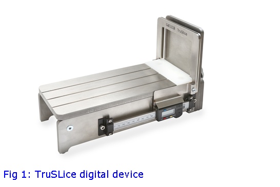
Whilst at my local delicatessen, I watched the assistant lift a large ham from the display counter, place it on a bacon slicer and cut several slices from the joint. He was unsure about how thin he could cut the slices but a glance at the display in front of me told me what I needed to know. Later I read about the bacon slicer and also about the guillotine, used during the French revolution as an instrument of death. What was evident was how successful these devices had been, the reason being they were perfectly constructed for their purpose. I started drawing some constructions and to ponder on how something that was not dissimilar to a guillotine or bacon slicer could be used for histological dissection, the biggest issue being how to ensure precision and accuracy. The best option appeared to be adapting the devices on which we already rely (i.e. the microtome) and adjust them for this purpose. With this in mind, I approached a commercial company (Cellpath) and discussed designs and formats. The literature in this area is not extensive and covers a wide spectrum of different approaches to tackling the needs of histological investigation. Some of the key factors that can affect accuracy and precision for histological dissection need to be appreciated and include:
- flat uniformity perpendicular to the specimen cutting face
- appropriate immobilisation of the tissue specimen during grossing
- good visualisation of the cutting tissue face
- sharp knives with associated grossing equipment fit for purpose
- grossing knife action
Designs were constructed that attempted to take into account these factors. What ensued were considerations of not just final designs but also the materials to be used in construction of these devices. Trials of two devices, TruSlice and TruSlice Digital, were performed. TruSlice relies on the insertion of reinforced plastic inserts with defined recessed depths of 2, 3, 4, 6 mm. The TruSlice Digital relies on the use of an attached micrometer. Both used a guillotine like construction with a knife plate configuration that ensured a perpendicular action of the blade. Preliminary trials were extremely positive with good recordings of accuracy and precision.
Following these encouraging results, I requested that we embark on a 5 site trial to determine if these initial findings were mirrored elsewhere and also to see how feedback could improve the devices.
The use of a micrometer (TruSlice Digital) to set tissue slice thickness is innovative and its use of a micrometer reminds us about how traditionally we have defined measurement and accuracy in the microtomy of histological sections from paraffin blocks. Clearly in developing these devices we have not reinvented the wheel but applied the concepts of well-trusted methodology to a different area of histological assessment.
There is a need, however, to compare and contrast the most promising devices in this area of investigation, simply to determine which will provide the best overall options for routine histological dissection. This is something that has not been performed to date. Owing to the complexity of, and variation in, tissue types dealt with in the modern histopathology laboratory, a single device that suits every need and eventuality with regard to dissection is perhaps an idealistic goal but that is not an excuse for not trying! As Thomas Huxley said: “Science is simply common sense at its best.”
References
Orchard G.E, Shams M, Nwokie T, Bulut.C, D’Amico C, Gabriel J, Ramji. Z, Georgaki A, Neichcial A, Shams F, Neesam H, Haine. N, Brewer C. Development of new and accurate measurement devices (TruSlice and TruSlice Digital) for use in histological dissection: an attempt to improve specimen dissection precision. Br .J. Biomed. Sci. 2015; 72(3): 140-5.
Orchard G.E, Shams M, Nwokie. T, Bulut C, Quaye CJ, Gabriel J, Ramji Z, Georgaki A, Watt. M, Cole S, Stewart K, McTaggart V, Padayachya S, Long A.M, Odgen A, Andrews C, Birchalls A, Neesam H, Haine N. TruSlice and TruSlice digital histological dissection devices, introducing an exciting development in providing improved accuracy and precision at the cut-up bench: Data from a five site trial throughout the United Kingdom. Br J Biomed Sci 2016 Oct; 73(4): 163-167.